
A modality is a method of which a diagnostic procedure is performed. The modalities are classified by the source of energy, sound, radio waves, visual light, and radiation, whether it's ionising or not. In the world of medical imaging, some examples of modalities are x-ray, computer tomography, magnetic resonance imaging, ultrasound, or nuclear medicine. First, let's discuss about modalities where ionising radiation is used and the source of radiation is a cathode ray tube.
- X-ray radiography
Here the source of radiation is ionising radiation where x-ray photons are produced by a cathode ray tube and shot into a patient. For the protocol, typically single static images are captured and separate views may be captured. The image type should contain static high-resolution images in greyscale. This would receive the DICOM modality tag of DX or CR. An example could be a chest x-ray or an x-ray of the hand where you take multiple views.
- Fluoroscopy
The source of radiation is a low dose ionising radiation. These are x-ray photons as well produced by the cathode ray tube. In contrast to an x-ray which takes spot views of high resolution images, the fluoro takes a continuous series of lower resolution images like a movie. The DICOM tag for fluoros are RF and an example of a fluoro could be a barium swallow study or a catheterisation.
- Computed tomography
In this type of imaging, consider an x-ray tube but instead of the static x-ray tube, it's moving in a circular motion or elliptical around the patient. There is a continuous acquisition of imaging as the patient is positioned in the gantry and the x-ray tube is rotating around the body acquiring thin transverse section of images. These slices can be reconstructed into multi-planar images. In other words, images with views from different angles. The DICOM tag for this modality is CT. An example would be a chest CT with or without contrast or a CT of the head.
Next, we'll discuss ionising radiation again but this time where the source of radiation is the patient.
In nuclear medicine, a patient has inhaled, consumed, or been injected with the radioactive isotope for diagnostic imaging. In most cases, the isotope emits gamma ray photons until it decays over x amount of time depending on the isotope. The patient can be imaged by single detectors where high resolution spot images can be taken similar to getting an x-ray or moving images can be captured called a SIN-A similar to a fluoro.
Additionally, they can be placed in a gantry where reconstructed SPECT imaging can be acquired similar to a CT. The important fact to note here is that the detectors don't admit radiation, they just capture the gamma photons that are being emitted from the patient. So, the DICOM tag is NM for this modality and examples are a lung VQ scan or a nuclear stress image.
- PET scan, positron emission tomography
PET falls under nuclear medicine, however, the isotope used here are positrons which are positively charged electrons producing a cyclotron. A PET scan can be combined with a CT. In an image, imagine a PET scan that can identify cancerous organs in a body by lighting it up a certain colour. When this image is overlaid with the CT, the physician can see a 3D image of the body from the CT plus organs which are lit up by the PET. The DICOM tag assigned to this modality is PT and examples are a full body FDG PET scan or a cardiac PET scan.
Next, we'll look at modalities that use non-ionising radiation such as sound waves, radio waves, or visible light.
- Magnetic Resonance Imaging or MRI
A patient is placed in a gantry with an extremely strong magnetic field. Radio frequency pulses switch on and off which align then relax the protons in the patient's body. The relaxation of the protons causes a release of radio waves which are captured by the detectors. These waves are plotted and reconstructed into multi-planar imaging. Now the modality DICOM tag for this is MR and examples are MR of the brain with or without contrast.
- Ultrasound
Ultrasound, otherwise known as sonogram, uses sound waves to capture images. Think of a submarine that produces sound waves outbound then captures the returning waves to produce an image. In medical sonography, a technologist presses a transducer against the patient's body. This handheld device both produces and receives sound waves. The returning sound waves are captured as either static or moving video images. The DICOM tag assigned to this is US. Examples of ultrasound include echocardiography of the heart and a venous doppler.
- Visible light
Photographic images of the body are taken by medical grade cameras. This includes high resolution images or movies. The DICOM tag assigned to this modality is VL and examples are microscopic slides of pathology, endoscopy videos, or dermatologic images of a patient's skin.
That just about sums it up as a very high-level overview of modalities.
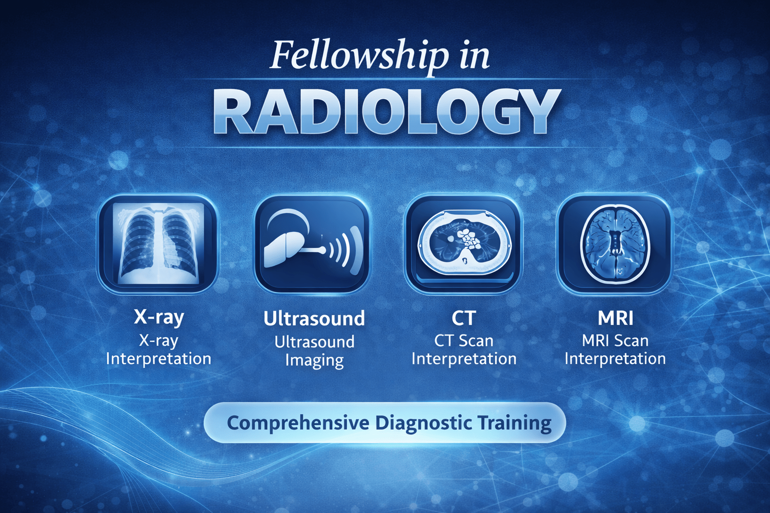
At Medline Academics, the Fellowship in Radiology is structured to assist doctors and imaging professionals in developing a solid grasp of both the theoretical and practical aspects of modern diagnostic imaging. The program is crafted to introduce learners to the core principles of various imaging techniques, including X-ray, CT scans, MRI, ultrasound, nuclear medicine, and PET scans. It goes beyond just listing these methods by explaining how each one operates, the situations in which they are most effectively used, and the process of capturing and analysing images within a clinical environment. The fellowship places significant importance on understanding the practical applications, the standard protocols followed, and the underlying physics that govern these imaging technologies.
In addition to radiology, this institute also provides a Fellowship in Embryology that emphasizes the science and accuracy behind assisted reproductive technologies. This fellowship offers comprehensive exposure to gamete manipulation, embryo growth, laboratory methodologies, quality assurance, and the ethical obligations of an embryology lab. Systematic academic learning is enhanced by clinical perspective, ensuring participants grasp not only how procedures are executed, but why each phase is vital to treatment results. Collectively, these fellowships demonstrate Medline Academics’ dedication to specialized, practice-focused medical education that aids professionals at every phase of their clinical careers.
Dr. Kamini Rao Hospitals, stands as a Top IVF Centre in Bangalore and reproductive health, grounded in the long-standing legacy of Dr. Kamini A. Rao, a Padma Shri award recipient and a trailblazer in the field of assisted reproductive medicine in India. The hospital provides an extensive array of fertility treatments, such as IVF, ICSI, IUI, and sophisticated reproductive diagnostics, all administered through customized treatment strategies that address the specific requirements of each couple. Equipped with cutting-edge technology and a team of specialists from various disciplines, including gynaecology, reproductive endocrinology, and embryology, the centre prioritizes comprehensive care that encompasses the reproductive health of both men and women. With over forty years of dedicated service and empathetic support, Dr. Kamini Rao Hospitals has gained the trust of couples in Bengaluru who are in search of informed and evidence-driven fertility solutions.